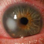
Previous
Next

SHARP & CRYSTAL CLEAR VISION 

What is Cornea

The outermost layer of the eye is called Cornea. It is one of the essential components of the human eye as it allows the light to enter into the eye for us to have vision. A cornea is generally 12mm in length and 11mm in height. The cornea also contributes towards around 70 percent of the focusing power of the eye. That is the main reason why it is imperative to take care of the corneas.

Refractive problems like myopia, hyperopia, and astigmatism are caused due to a change in the shape of the cornea.

joh_cornea2

Our Cornea Service includes a full-time staff of attending surgeons. Each patient is carefully examined by our specialist and then given an individualized treatment plan, based on the latest and best therapies. We are dedicated to improving vision, preventing corneal and external diseases of the eye, and providing the best patient care available.

ulcera-corneal1

Corneal Disease

The cornea is your eye's clear , protective outer layer. Along with the sclera ( the white of your eye), it serves as a barrier against dirt, germs and other things that can cause damage.
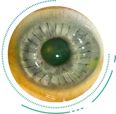
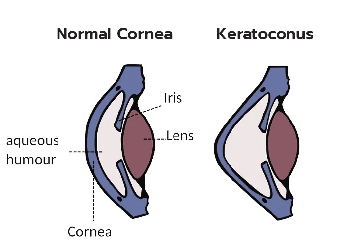
4d326a2f-5aef-4a4f-a642-c7c5642b2a17

Anterior Segment

The anterior segment of the eye encompasses the cornea, iris, lens and aqueous humor, which provides nutrients to the avascular cornea and lens.
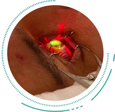
preparing-patient-for-laser-eye-surgery-1157972284-81a2d6bff7574014b3dc9ed8c214e681

Refractive Surgery

Refractive lens exchange surgery involves carefully removing the natural lens in the eye through a small incision and replacing it with an artificial intraocular lens implant.

You May Need Cornea Opinion If You Have Any Of These Symptoms

SAVE YOUR SIGHT FOR THE FUTURE

What Happens In Keratoconus

The Cornea is the front part of the eye lens, made of collagen fibers, which makes the light fall onto the retina. In a Keratoconus Eye, the layers of the cornea start thinning out, moreso in the central area. This thinning out causes the cornea to become weak and change its shape from a dome-like structure to a cone-like structure which leads to induction of high Astigmatism.

This thinning also causes the cornea of the Keratoconus Eye to become irregular in shape which in turn prevents all the light rays entering our eyes to focus accurately onto the Retina. These two factors cause a drop in the patient’s vision.

At late stages of the disease, the cornea becomes extremely thin and is unable to withstand the pressure from within the eyes. This leads to small tears in the Cornea and accumulation of fluid in its layers leading to a condition called ‘Acute Hydrops’. This causes a sudden loss of vision accompanied by pain, watering and redness. Keratoconus Treatment in early stages and advanced stages of this disease vary significantly.

HOW DO I KNOW IF I HAVE KERATOCONUS?

Symptoms

Keratoconus usually affects teenagers and young adults

Frequent change of glasses
Vision not correcting even with glasses
Headache

Causes Of Keratoconus

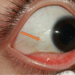
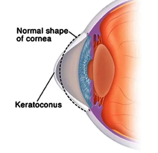
Although there is no proven cause for Keratoconus, numerous risk factors have been identified which can trigger the progress of the condition.

  • Right Arrow
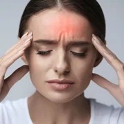
Vigorous Eye Rubbing, especially in childhood [due to allergic causes] is one of the major identified risk factors of Keratoconus.

Some of the other risk factors include family history, age, race and certain genetic conditions such as down syndrome.

HEALTHY EYES FOR CLEAR VISION

Keratoconus Treatment

Keratoconus Treatment cannot reverse the thinning of the Cornea. However, the basis of Keratoconus Eye Treatment is to prevent the further progression of the disease so as to reduce the extent of vision loss caused by the disease. Early Keratoconus Treatment is always advocated to prevent adverse complications. Modern Keratoconus Treatment modalities are safe and effective with a proven track record.


Riboflavin Assisted Corneal Collagen Cross-Linking [C3R]

Corneal Cross Linking is a type of Cornea Surgery and is the most commonly recommended type of Keratoconus Treatment to strengthen the Corneal Eye Layers. This Cross-linking Treatment involves the use of Riboflavin drops which soak into the Cornea. UV radiation is then applied to stimulate the corneal layers to form strong chemical bonds which improve the strength of these layers.

The C3R Keratoconus Surgery thus prevents the cornea from weakening and bulging further. C3R procedure if done at an early stage can safely help in ensuring that vision loss does not progress. Keratoconus Eye Surgery cannot be done at later stages of the disease when the cornea becomes extremely thin or when the integrity of the cornea has been compromised [such as due to scarring or acute hydrops].

In cases of extremely thin Corneas, Keratoconus Treatment using Corneal Cross Linking can still be done with some modifications such as Hypotonic C3R, contact lens assisted C3R, etc. In case of scarred corneas or compromised corneas, C3R Surgery would not be effective and the Cornea Surgery of choice would be to perform a Corneal Transplant.

All patients require Visual Rehabilitation after Cross Linking Surgery to complete the Keratoconus Treatment as it DOES NOT REVERSE the Corneal Astigmatism. Visual Rehabilitation is usually in the form of spectacles [in very early stages of the disease] or in the form of specialized contact lenses such as rigid gas permeable lenses[RGP’s], Rose-K contact lenses, Scleral or Mini-Scleral Lenses.

Keratoplasty: Lamellar(DALK) or Penetrating [Corneal Transplantation]

Keratoplasty is a Cornea Surgery where a donor healthy cornea is grafted in the place of a diseased host cornea. Keratoplasty for a Keratoconus Eye is only reserved for advanced cases where C3R surgery is contraindicated or in cases of corneal scarring or corneal compromise. The diseased cornea of the patient is removed out and the donor cornea is grafted in its place. Depending on the extent of the Keratoconus and the status of the Cornea, Corneal Transplantation for Keratoconus can be:

Penetrating Keratoplasty:
In this Keratoconus Treatment, all the layers of the cornea layer (full thickness) are replaced with the donor’s cornea. Penetrating Keratoplasty Cornea Surgery is usually recommended when the corneal layers are extensively affected, including its water tightness and transparency.

Lamellar Keratoplasty:
[Partial thickness Corneal Transplantation Cornea Surgery] For a Keratoconus Eye with extreme corneal thinning but whose water-tightness is still present, a partial thickness grafting of only the front layers of the cornea is performed as a method of Corneal Cross Linking Keratoconus Treatment. Lamellar Keratoplasty has the advantage of having a lower graft rejection and failure rate as compared to the Penetrating Keratoplasty Procedure of Keratoconus Treatment.

MEET OUR CORNEA DOCTOR

DR. M.K. Sanghi

MBBS & MS (MAMC, DELHI), DOMS, MAMS (VIENNA)

Dr M.K. Sanghi is the Director & Senior Phaco, Refractive & Ant. Segment and Glaucoma Surgeon at Shekhar Eye Centre.

He had graduated from PGIMS Rohtak and completed his medical and ophthalmology training from the prestigious Maulana Azad Medical College in Delhi.

After a short stint of working in UK & Middle East, he has been practicing in Delhi since 1987.

Monday - Saturday

10:00 am to 06:00 pm

Contact us

011-35614362 ,+91 93 122 65648

E-mail

shekhareyecentre@gmail.com